Role of the Circulatory System:
- A network of blood vessels that distributes gases, nutrients, waster products, and cell products (hormones) and distributes heat
- Unicellular organisms such as bacteria do not need a circulatory system since they are in direct contract with their environment and activity can take place across the cell membrane
- Larger organisms need a circulatory system in order to transport materials throughout the organism’s cells
Circulatory System Requirement:
- Require a fluid to transport materials
- Network if tubes for fluid
- Pump to push fluids through tubes
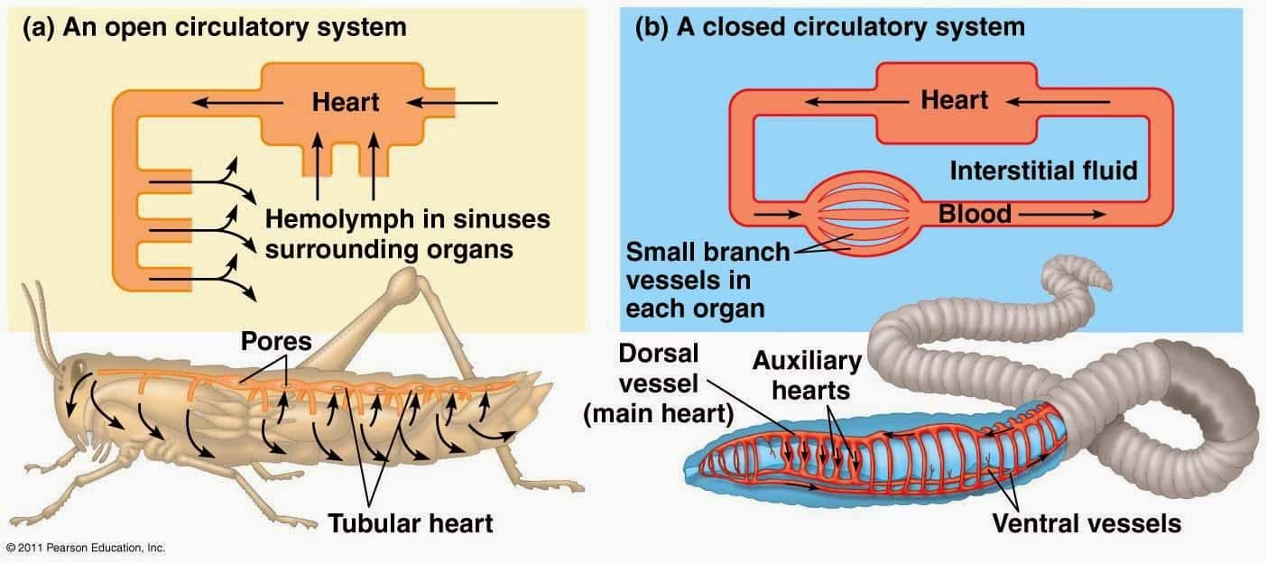
Open Circulatory System:
- Appears in invertebrates such as worms and insects
- A circulating fluid called hemolymph is pumped into an interconnected system of body cavities (sinuses) where the body cells are surrounded by fluid
- Lower metabolic rates and do not need to eat often
Closed Circulatory System:
- All vertebrates and some invertebrates have a closed circulatory system
- The fluid remains in the network of blood vessels
- The blood vessels and body tissues are kept separate
Blood:
- Blood consists of cells suspended in a liquid called plasma
- The function is to transport nutrients, gases, cell products and wastes to and from cells
Plasma:
- Plasma is a protein-rich liquid that blood cells and platelets are suspended into
- 90% of plasma is water and it contains dissolved solutes (sodium, calcium) as well as nutrients, respiratory gases, metabolic wastes and hormones
- Plasma proteins control blood pH
- Albumin controls the amount of water that enters the bloodstream
- Globulins transport insoluble lipids, cholesterol, mineral and fat-soluble vitamins
- Antibodies guard against different microorganisms
- Fibrinogen is involved in blood clotting
Cells:
- Consist of red blood cells, white blood cells, and platelets
- Always being replaced
- Can specialize into different cells
Red Blood Cells:
- Also called erythrocytes
- 90% of the cells in your body
- Thinner in the center then at the edge, increases the surface area
- The function is to deliver oxygen and some carbon dioxide
- Hemoglobin is a protein with an iron atom that binds oxygen and carbon gases
- Only live for 120 days and are removed by the liver and spleen when they die
- EPO hormone in the kidneys stimulates red blood cells production
- No nuclei in mammals, and lacks mitochondria to make ATP
White Blood Cells:
- Also called leukocytes
- Less than 1% of blood cells are white blood cells
- The function is to protect the body from harmful bacterial viruses and other foreign invaders
- White blood cells have a nucleus
- The number of white blood cells increases when someone is fighting an infection
- 5 types of white blood cells-3 are granular, 2 are agranular
- Granular contain chemicals to attack foreign material and microorganisms. Include: neutrophils, basophils, eosinophils
- Agranular release enzymes to destroy foreign objects. Include: monocytes, lymphocytes
Platelets:
- Have no nuclei
- Involved in blood clotting and are small pieces that have been broken off from the cells in bone marrow
- Broken blood vessels with rough edges with break platelets which release chemicals that help platelets stick together to form a plug. The platelets then form a clot which clogs the tear in the blood vessels to prevent the loss of blood cells
Blood Types:
- Blood types are determined by the presence or absence of different sugar molecules attached to the cell membrane of red blood cells
- These sugars are called antigens
- Four blood types: type A- A antigen is present, type B- B antigen is present, type AB- both antigens are present, type O- no antigen is present
Antigens:
- An antigen is any molecule to which the immune system can respond
- If the immune system finds an abnormal antigen it will begin to fight against it. However, normal antigens found on the body’s own cells are known as self-antigens and not usually attacked
- The antigens in the red blood cells are what determines a person’s blood type. There are two antigens, A and B
Blood Transfusions:
- May be required to treat a medical problem
- When individuals receive the wrong blood type, antigens are considered foreign objects to the body, which causes the immune system to react, producing antibodies to attach to antigens.
- This causes agglutination-blood cells that clump together blocking circulation and oxygen supply
- Type 0 blood is considered a Universal Donor therefore, can donate to all blood types
- Type AB can receive from all, therefore Universal Acceptor
| Student | A | B | C | D |
| Blood Type | AB- | O- | B+ | A+ |
| Can Donate To | AB | A, B, AB, O | B, AB | A, AB |
| Can Receive From | A, B, AB, O | O | B, O | A, O |
The Rhesus Factor:
- Inherited protein found on the membrane of the red blood cells affects the compatibility of the blood types
- It is an antigen that produces an antibody reaction
- Rhesus antigen is present in 85% of the red blood cells, the remaining 15% do not have it
- Rh+ (positive)= have the rhesus factor antigen on red blood cells, only donate to Rh+
- Rh- (negative)= do not have the rhesus factor on red blood cells, can donate to anyone
Blood Vessels:
- A complex network of tubes that branch and re-branch to distribute blood and its
 contents to the body cells
 - The largest are 3cm wide, and the smallest can still have cells pass through in a single file
- 5 main types include: arteries, arterioles, capillaries, venules, veins
Arteries:
- Carry blood away from the heart toward body tissues
- Usually, the blood has oxygen
- Oxygenated blood is pumped out of the heart through the aorta
- When the heart squeezes in, it pushes blood into the major arteries
- The artery walls expand to allow the increased blood volume and pressure; this is how you feel your pulse
- When the heart finally relaxed, the artery walls return to their original size and push the blood further along the blood vessels
Arterioles:
- Smaller arteries
- Size is controlled by nerve signals from the brain
- Vasodilation: when the arterioles dilate to increase blood flow (when you are hot, they dilate to increase blood flow, so you can lose heat)
- Vasoconstriction: when the arterioles contract to reduce blood flow to the skin to prevent loss of heat to the outside environment
Capillaries:
- Smaller blood vessels are formed when arterioles reach the tissue
- A capillary network is an extensive network of blood vessels that supply oxygen and nutrients to every cell throughout the body tissue
- Walls are only 1 cell thick so they allow diffusion of materials from the blood vessels to the cells
- pre-capillary sphincter muscles can control blood flow into a capillary, when blood is needed the sphincter relaxes, and when blood is not needed the sphincter contracts
Veins & Venules:
- Venules are small veins, that merge to form veins
- Veins carry deoxygenated blood toward the heart, contains carbon and other waste
- Veins contain one-way valves that are in between skeletal muscles and allow blood to flow in one direction only. When the muscles contract, blood is forced through the vein back to the heart
- Varicose veins are damaged venous valves that accumulate blood in the veins resulting in the formation of a bulge
The Heart
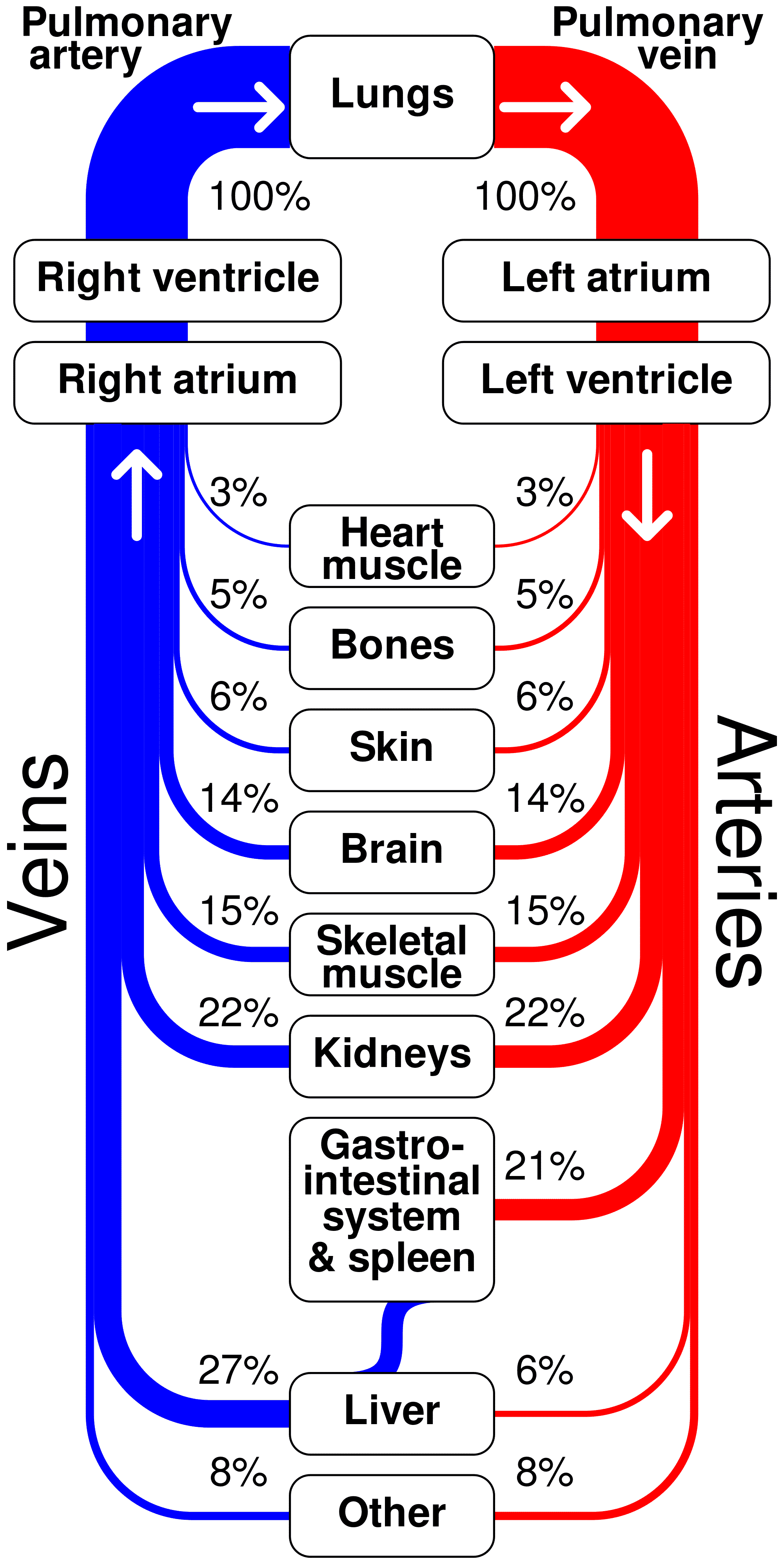
- As mammals, we have a 2-circuit system
- Pulmonary Circuit: Blood is pumped to and from the lungs to get oxygenated
- Systemic Circuit: Blood is pumped to and from the body to deliver oxygen and returns deoxygenated blood
Heart Structure
- The heart is made up of cardiac muscle (which is a myogenic muscle)
- The heart is located in the middle of the chest, which is under the breastbone *called the sternum
- Surrounded by the pericardium, a two-layered fluid-filled membrane and its goal is to stop the friction between the heart and other tissues and organs as the heartbeats
- The heart spontaneously contracts and relaxes without the nervous systems controls
Different Chambers of the Heart
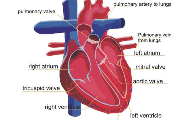
- A wall of muscle called the septum separates the heart into 2 parts, where each part has a ventricle and atrium
Atria
- The atria are located at the top of the heart; they receive blood to be further pumped into the ventricles
- The right atrium receives blood from the body
- The left atrium receives blood from the lungs
- The muscle around the atria is thin because it pumps blood over short distances (only to the ventricle)
Ventricles
- Ventricles, which are located at the bottom of the heart, pump blood through the pulmonary and systemic circuits
- Thicker walls of muscle surround ventricles because they have to pump blood further than the atria
- The left ventricle has a thicker coating of muscle than the right because it needs to pump blood to the whole body while the right ventricle only needs to pump blood to the lungs
Circulation
- Four valves ensure that blood flows in the right direction within the heart. Two semilunar valves and two atrioventricular valves
- Semilunar valves include the pulmonary valve and aortic valve; they prevent blood from flowing back into the ventricles when the ventricles relax
- Atrioventricular valves include the tricuspid and bicuspid valves; they prevent blood from flowing back into the atria from the ventricles
Heart Rate
- Heart rate is the number of times a heart beats each minute.
- The pulse us the stretching of the arteries as they receive the pressure exerted by the blood
- A cardiac cycle (a complete heartbeat) takes normally 0.8 s.
- During a heartbeat, the heart pumps twice, 2 contractions: 1 atrial and 1 ventricular.
Heart Rhythm
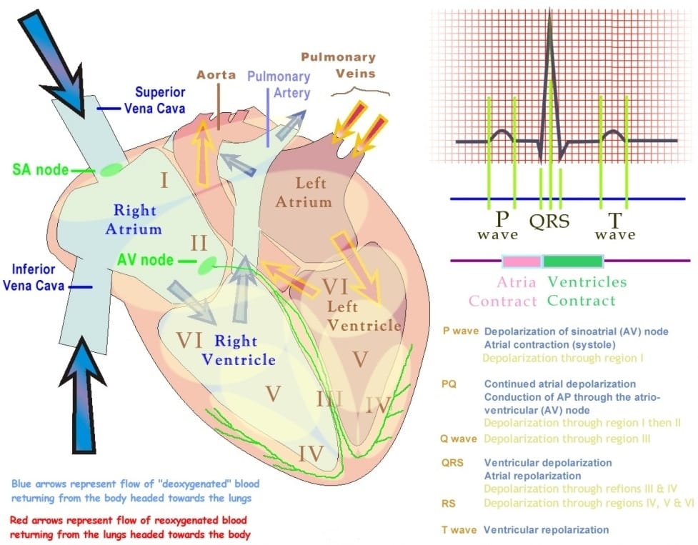
- The heartbeat is triggered by the SA Node (Sino-Atrial Node), also known as the pacemaker
- The SA nodes send “electrical signals” to both atria causing them to contract simultaneously. Allowing blood to be pushed out of the atria and into the ventricles
- At the same time it sends an electrical impulse to the Atrioventricular Node (AV node), the AV node sends signals to fibers called the Purkinje fibers; this causes both ventricles to contract, pushing blood out of the ventricles into the arteries
Cardiac Cycle
- There are two phases to the heartbeat (cardiac cycle), the diastolic phase and the systolic phase. The diastole fills the heart with blood while relaxing and the systole has to do with the emptying of the heart during contraction
- Stages of the cycle:
- Stage 1 Diastole: The heart is relaxed and the atria begin to fill with blood
- Stage 2 Diastole: As atria fill, the pressure opens the atrioventricular valves and blood enters the ventricles
- Stage 3 Diastole: Muscular walls of the atria contract to fully push all the blood from the atria to the ventricles
- Stage 4 Systole: Once ventricles are filled, they contract; forcing the atrioventricular valves to close and the semilunar valves remain closed
- Stage 5 Systole: The ventricles fully contract, the semilunar valves are forced open and blood flows into the pulmonary and aortic arteries

Heart Sounds
- The lubb-DUBB sound that is heard from the stethoscope is made by the closing of the atrioventricular valves as the ventricles begin to contract (lubb) and the DUBB sound occurs when the ventricles relax and the semilunar valves snap shut
Control of Heart Rate
- Though the SA node sets the tempo for the heartbeat, the brain and hormones influence the frequency of the heartbeats
- Two sets of nerves, “sympathetic” and “parasympathetic” nerves from the brain connect to the SA node
- During stress, the sympathetic nervous system increases heart rate.
- During times of relaxation, the parasympathetic nervous system functions to decrease heart rate
- Hormones and body activity also affects heart rate
- Athletes have a lower resting heart rate than the average person because their heart is trained to pump more blood in one contraction so there is no need to pump as many times
ECG – Electrocardiogram
- The cardiac cycle can be monitored using an electrocardiograph, which measures electric signals and produces an ECG
- PQRST model:
- P: Contraction of atria
- QRS: Contraction of ventricles
- T: Recovery period; heart is preparing for the next contraction
Heart Murmur
- Defect is one or more valves causing a hissing sound of blood squirting back through the valves
- Problematic because this means not all blood is flowing forward, therefore less oxygen is delivered to the body, making the heart beat faster to make up for lost oxygen
Blood Pressure
- Blood pressure is the pressure exerted by the blood on the walls of the vessel
- When blood vessels are stretched to the limit, the increase in blood pressure can cause major health risks.
- Blood pressure can be measured with an instrument called a sphygmomanometer
- The readings on this instrument have two readings
– Systolic pressure: the pressure when the heart contracts
– Diastolic pressure: the pressure when the heart is at rest (relaxed)
- Normal blood pressure is 120/80 (systolic/diastolic)
- Some factors that affect blood pressure include:
- Temperature
- Diameter of blood vessels
- Smoking
- Diet
- Age
- Stress
- Constantly high blood pressure is called hypertension, can be due to a variety of factors, one being a high sodium diet
Arteriosclerosis
- Arteriosclerosis is the hardening of the arteries over time, too much pressure can cause the arteries to lose elasticity and harden
- Symptoms of arteriosclerosis include high blood pressure, poor circulation, recurrent kidney infections, and heart attacks
- Arteriosclerosis can branches off into atherosclerosis and coronary heart disease
Atherosclerosis
- Hardening of the artery walls caused by a build-up of plaque in the artery walls
- The plaque narrows the passage and creates a clog, which is dangerous because it can cut off blood flow to an organ, this can happen to any organ, not just the heart
Coronary Artery Disease (CAD)
- This disease occurs when atherosclerosis happens in the coronary arteries, which supply the heart with blood
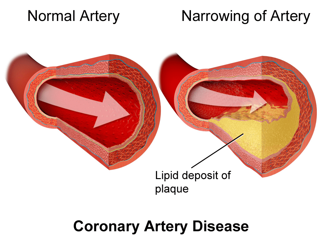
- CAD can happen due to many factors such as high blood pressure, high cholesterol, being overweight, smoking, gender, smoking, age or genetics
- A common symptom of CAD is angina; this is pain in the left chest, shoulder or neck, due to the insufficient blood supply to the cardiac muscles. It is treated with nitroglycerin, which helps pick up blood flow
Heart Attack
- Myocardial infarction (heart attack) is the death of an area of the heart muscle due to oxygen deprivation. Plaque build up in the coronary artery often ruptures, causing a blood clot, which reduces blood flow and less blood means less oxygen to the heart
- After 20-40 minutes, the heart cells begin dying due to the lack of oxygen; they will continue dying unless treated. This becomes fatal if a large area of the heart is affected
- Symptoms of a heart attack include chest pain, difficulty breathing, pain in the arm back or jaw, nausea or vomiting and others
- Treating heart disease can include lifestyle changes like exercise, eating healthy or quitting smoking
- Medication can be used to reduce build up of plaque
- Angioplasty can be performed, which opens a blocked artery by inflating a balloon at the point of blockage. A stent may be placed to ensure the artery stays open
- Bypass surgery can be done, which re routes or ‘bypasses’ the blood around the clogged arteries to improve blood flow
Stroke
- Strokes occur when blood vessels leading to the brain are damages or blocked and prevent blood and oxygen from getting to the brain, brain cells then die.
- Causes of stroke include atherosclerosis in the arteries of the brain or neck, blood clot in a blood vessel in the brain or cerebral hemorrhage (rapid loss of blood)
- Stroke (depending on how severe) can cause paralysis, vision problems, memory loss, difficulty breathing or loss of balance.
