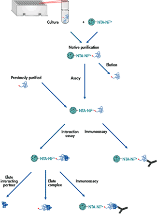
It is often necessary to obtain a preparation containing protein molecules of only a single type (a “pure protein”); eg. for medical use or scientific study. With such preparations, one may determine 3D structure, binding affinities, or determine the amino acid sequence. Obtaining a pure protein is generally challenging because there are thousands of proteins inside a cell as well as DNA, RNA, etc., all of which must be removed to leave a homogeneous sample containing only the protein of interest. In many cases, the advent of genetic engineering has simplified the problem by allowing amplification of the gene encoding the protein and the addition of “tags” to the protein sequence. Regardless, the protein must still be “fished out” of a complex mixture.
The purification procedure involves a number of steps, depending on the properties of the desired protein. Generally, these steps involve cell breakage, centrifugation for initial fractionation, then some form of column chromatography to separate proteins from one another. Small samples of the preparation are monitored at different stages by SDS (sodium dodecyl sulfate) polyacrylamide gel electrophoresis to determine the number of proteins present. Measurement of the protein of interest by activity or other means allows determination of the degree of purity.
Proteins differ from one another in their sequence and therefore in their properties of net charge, size, shape, hydrophobicity and binding affinity. Various types of liquid chromatography take advantage of these differences to separate proteins. Gel filtration separates on the basis of size, ion exchange chromatography on the basis of net charge, and affinity chromatography on the basis of binding affinities. How these methods work will be discussed in class.
Once pure protein is obtained, it is sometimes possible to determine its 3D structure by using either X-ray diffraction or nuclear magnetic resonance (NMR) techniques. Both methods have limitations and take a great deal of work to succeed, but they have led to detailed knowledge of more than 14,000 proteins so far.
In particular, X-ray diffraction requires making crystals of the protein, which can be a very difficult and time-consuming process. Once crystals are created, a beam of X-rays is passed through the crystal and some are deflected by the electrons present in the protein. This gives a pattern of spots (a diffraction pattern) that is then used to calculate the positions of the deflecting atoms, giving an exact 3 dimensional picture of the protein in the crystal.


