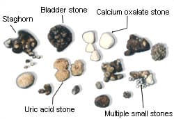
What are Kidney Stones?
- Small, hard mineral and acid salt deposits that form inside the kidneys.
- Often occur when the urine becomes concentrated, allowing minerals to crystallize and stick together.
- May affect any part of the urinary tract — from your kidneys to your bladder
- Stones are classified by their location in the urinary system and their crystal composition.
Statistics
- The lifetime incidence of kidney stones is nearly 13% in men and 7% in women.
- Once an individual has formed a stone, the likelihood of recurrence is 50% or greater at 5 years and up to 80% at 10 years.
- Kidney stones can develop at any time, although they are most common among adults over the age of 30.
Types
Four major types:
- Calcium stones: “Most common”. Occur in two major forms: Calcium oxalate stones which may be caused by high
 calcium and high oxalate excretion and calcium phosphate stones which are caused by the combination of high urine calcium/alkaline urine, meaning the urine has a high pH.
 - Uric acid stones: Form when urine is persistently acidic. A diet rich in purines (found in meats, fish, & shellfish) may increase uric acid in urine, which if becomes concentrated, can settle and form a stone.
- Struvite stones: Result from kidney infections. “Staghorn Stone”
- Cystine stones: Result from a genetic disorder that causes cystine to leak through the kidneys and into the urine, forming crystals that tend to accumulate into stones.
A Brief History
The first known stones were discovered in 1901 inside the bladder of a 5000 year old Egyptian mummy!
Surgery to treat stones was first described by an ancient Indian surgeon named Sushruta in the 8th century B.C. He provided detailed information on urinary stones, urinary anatomy, and surgery for stones in his writings, compiled as the Sushruta Samhita.
Hippocrates (4th century B.C.) specifically mentioned kidney stones in his Hippocratic Oath which is historically taken by newly trained physicians. “I will not use the knife, not even on sufferers from stone, but will withdraw in favor of such men as are engaged in this work.”
In the middle ages, surgeons called “lithotomists” traveled around Europe with special tables called “lithotomy tables” on which they placed patients to cut out stones. The term “lithotomy position” is still used to refer to placing a patient in a position similar to a woman in childbirth.
What Causes It?
- Dehydration: (Most common cause) When you don’t drink enough water the salts/minerals & other substances in the urine can stick together to form a stone.
- Animal proteins: (Red meat, poultry, eggs, & seafood) To much boosts the level of uric acid and could lead to kidney stones. High protein diets also reduce levels of citrate, the chemical in urine that helps prevent stones from forming.
- Medical conditions: Such as gout and Crohn’s disease.
- Genetics: May occur in family members over several generations.
- Parathyroid glands: Kidney stones form, in rare cases, because the parathyroid glands produce too much of a hormone leading to higher calcium levels and possibly calcium kidney stones.
How Is It Diagnosed?
If your doctor suspects you have a kidney stone, you may have diagnostic tests and procedures, such as:
- Blood testing: Blood tests may reveal too much calcium or uric acid in your blood.
- Urine testing: The 24-hour urine collection test may show that you’re excreting too many stone-forming minerals or too few stone-preventing substances.
- Imaging: These tests may show kidney stones in your urinary tract. (X-rays, CT scans, and ultrasounds.)
- Analysis of passed stones: You may be asked to urinate through a strainer to catch stones that you pass. Lab analysis will reveal the makeup of your kidney stones and this information will be used to determine what’s causing your kidney stones and to form a plan to prevent more.
Signs & Symptoms
A kidney stone may not cause symptoms until it starts moving around inside the kidney or passes into your ureter. At that point, you may experience these signs and symptoms:
- Severe pain in the side and back below the ribs or in the lower abdomen and groin
- Pain that comes in waves and fluctuates in intensity
- Pain on urination
- Pink, red or brown urine that may be cloudy or foul-smelling
- Nausea and vomiting
- Persistent need to urinate or only going in small amounts
- Fever and chills if an infection is present
Disease Progression
If a kidney stone is left alone, it will pass naturally through the body within a couple days or weeks, given it is the adequate size. ( less than 4mm in diameter) If it is 6mm or larger, it may need treatment to be removed another way. (next slide)
An untreated obstructing stone that causes persistent blockage can eventually cause atrophy in a kidney, resulting in a dilated, thinned out kidney with minimal function. Thankfully, because most stones are associated with significant amounts of pain, most patients seek treatment before permanent damage occurs. However, in cases where patients have “silent” stones with little or no pain, long term obstruction can lead to kidney damage.
Infections may also result leading to UTI’s.
Treatment
Treatments to remove large stones include:
Extracorporeal Shock Wave Lithotripsy (ESWL) – (Most common) Ultrasound is used to pinpoint kidney stone. Ultrasound  shock waves are then sent to the stone from a machine to break it into smaller pieces, so it can be naturally passed.
Ureteroscopy – A long, thin telescope (ureteroscope) is passed through your urethra and into your bladder while under general anesthetics. It’s then passed up the ureter to where the stone is stuck. The surgeon may either try to remove the stone or use a laser to break it up.
Percutaneous Nephrolithotomy (PCNL) – An alternative for larger stones or if ESWL isn’t suitable (ex. Obese). A small incision is made in your back while your under anesthetics and a thin telescopic instrument (nephroscope) is passed through it and into the kidney. The stone is either pulled out or broken into smaller pieces using a laser or pneumatic energy.
Open surgery – (Rare) Only used for large stones or abnormal anatomy. During open surgery, an incision will be made in your back so that your surgeon is able to access your ureter and kidney and the stone is then removed by hand.
References
Cause. (2014, November 14). Kidney Stones Health Center. Retrieved September 25, 2016, from http://www.webmd.com/kidney-stones/tc/kidney-stones-cause
History of kidney stones. (2015). Retrieved September 25, 2016, from http://www.kidneystoners.org/information/history-of-stones/
(2015, February 26). Symptoms. Diseases and Conditions Kidney Stones. Retrieved October 1, 2016, from http://www.mayoclinic.org/diseases-conditions/kidney-stones/basics/symptoms/con-20024829
Mayo Clinic Staff. (2015, February 26). Tests and diagnosis. Healthy Harvard Publications Diseases and Conditions Kidney Stones. Retrieved September 25, 2016, from http://www.mayoclinic.org/diseases-conditions/kidney-stones/basics/tests-diagnosis/con-20024829
Pendick, D. (2013, October 04). 5 steps for preventing kidney stones. Healthy Harvard Publications. Retrieved September 25, 2016, from http://www.health.harvard.edu/blog/5-steps-for-preventing-kidney-stones-201310046721
Treating Kidney Stones. (2016, June 15). NHS Choices. Retrieved October 1, 2016, from http://www.nhs.uk/Conditions/Kidney-stones/Pages/Treatment.aspx#
Urology. (2013, June 23). UW Health. Retrieved October 18, 2016, from http://www.uwhealth.org/urology/how-common-are-kidney-stones/11208


It was very helpful! I am also looking for herbal treatment of kidney stones.
My son has a small kidney stone, and we are trying to decide the best treatment for him. I like the idea of using the extracorperal shock wave lithotripsy, since that is the best way to help it pass naturally way.